A. Anteroseptal myocardial infarction – Explanation
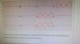
We can note that there is ST elevation in lead V2, V3, V4 (circled in red). This ECG shows a classical
example of an anterior myocardial infarction.
Those ECG findings are more than enough to answer the questions in the exam and it is highly unlikely
that you would need to know more than that.
For those who want to go into more details (probably noted needed for the exam), one can notice the
following on this ECG:
- Q waves are present in the septal leads (V1-2)
- Note the subtle ST elevation in I, aVL and V5, with reciprocal ST depression in lead III
- There are hyperacute (peaked) T waves in V2, V3 and V4
These features indicate an anteroseptal STEMI
ECG CHANGES IN MYOCARDIAL INFARCTION AND CORONARY ARTERIES
| Area of Infarct | ECG Changes | Coronary Artery | |
| Most commonly asked | Anteroseptal Inferior Latera |
V1-V4 II, III, aVF I, aVL +/- V5-6 I, aVL, V4-6 |
Left anterior descending (LAD) Right coronary (RCA) Left circumflex Left anterior descending (LAD) or left circumflex |
| Less commonly asked | Posterior Posterior |
Tall R waves V1-2 Also note the reciprocal ST-segment depression in the anterior chest leads |
Usually left circumflex, also right coronary |