C. Placenta praevia Explanation
praevia.
In the UK, it is common to have an ultrasound scan during the first trimester and again
around 20 weeks for an anomaly scan which the sonongrapher would formally report
the position of the placenta. In this stem, she did not have previous scans and so she
may very well have a low lying placenta.
The painless bleeding seen in placenta praevia may occur without warning or after
intercourse. This is one reason obstetricians advise women with placenta praevia not to
have intercourse.
From her observations, one can see that it is maternal blood that is being lost which is
consistent with placenta praevia. If this was the case of vasa praevia, it would be foetal
blood that would be lost and maternal observations would remain normal.
Placental abruption presents with sudden onset abdominal pain +/- vaginal bleeding. It
is also commonly associated with CTG abnormalities.
Placenta accreta is less common than placenta praevia and are seen commonly in the
presence of a uterine scar which alows the placenta to attach to the myometrium.
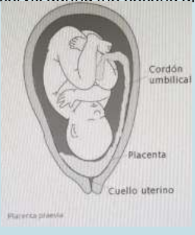
PLACENTA PRAEVIA
Placenta praevia describes a placenta lying wholly or partly in the lower
uterinesegment. This is common early in the pregnancy, but is most often not associated
with bleeding.
The key clinical feature is painless bleeding after 24 weeks of gestation.
Risk factors
• previous placenta praevia
• multiple pregnancies
Clinical features
• Painless vaginal bleed
• uterus not tender
• lie and presentation may be abnormal
• fetal heart usually normal
The painless late-pregnancy bleeding may occur during rest or activity, suddenlyand
without warning. It may be preceded by trauma, coitus, or pelvic examination. A digital
vaginal examination should not be performed. However, a speculum or a transvaginal
probe can safely be used in placenta praevia.
Diagnosis
This is based on the presence of painless late-trimester vaginal bleeding with
anobstetric ultrasound showing placental implantation over the lower uterine segment.
A transvaginal ultrasound is preferred over abdominal ultrasound for detection
ofplacenta praevia.
In the UK, most low-lying placentas are detected at the routine anomaly scans (around
20 weeks gestation). This is done transabdominally. If the placenta extends over the
internal cervical os, they are offered another transabdominal scan at 32 weeks. If the
position of the placenta is still unclear using a transabdominal scan, a transvaginal scan
is offered.
Around 5% will have low-lying placenta when scanned at 16-20 weeks gestation
however the incidence at delivery is only 0.5%, therefore most placentas rise away from
cervix during the second and third trimester.