E. Spontaneous pneumothorax – Explanation
mediastinal shift to the left.
The right lung is completely compressed and the trachea is pushed to the left. It
is obvious that there are no lung markings on the right field. You will not be
asked complicated X-rays in the exam, but life threatening X-rays like this where
it is easy to spot should not be missed in the exam and also in practice.
An urgent chest drain would be required to alleviate her symptoms and to
prevent worsening of the mediastinal shift.
Although she is likely to be an undiagnosed COPD patient given the history of
her smoking, the more important diagnosis at the moment is the right
pneumothorax. The undiagnosed COPD that this patient has is likely the cause
of her secondary spontaneous pneumothorax.
Remember, if it is clinically clear that you have a patient with tension
pneumothorax, do not request an X-ray just yet, treat the patient!
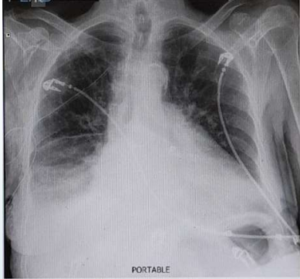
This chest X-ray is taken after the chest drain insertion showing the right sided
intercostal drain in situ with good reexpansion of the right lung with no evidence
of residual pneumothorax. If you look carefully, a consolication is seen on the
right lower lobe which could account for her symptoms of a lower respiratory tract
infection.